MORPHOLOGY OF MALARIAL PARASITES
WHAT IS MALARIA?
•Malaria is a disease caused by parasite that enters the
blood.
•This parasite is a protozoan called plasmodium.
•3 to 700 million people get malaria each year, but only
kills 1 to 2 million
•40% of the worlds population lives in malaria zones
•Malaria zones are: Africa, India, Middle East,
Southeast Asia, Central and South America, Eastern Europe, and the South
Pacific
MALARIA PARASITE (PLASMODIUM)
•Pathogen of malaria
Plasmodium vivax
Plasmodium falciparum
Plasmodium malariae
Plasmodium ovale
•P.vivax ; P.falciparum are more common
•Plasmodium is a wide distribution in many tropical or
subtropical regions of the world
MALARIA – VECTOR
 |
| Anopheles stephensii - common vector in India |
 |
| Plasmodium vivax - Various stages seen microscopically |
Plasmodium vivax
Early
trophozoite (ring form)

- Red nucleus on the ring, with light blue cytoplasm
- Infected RBCs are enlarged
Late trophozoite

It is irregular shape like ameboid form with pseudopodia; within cytoplasm ,brown pigment granules (malarial pigment---haemozoin) appear.
Immature schizont

Oval in shape, nucleus divided into 2-4 or more, malarial pigment begins to
concentrate in a mass.
Mature schizont

Nucleus
divided into 12-24; and cytoplasm also divided , each nucleus surrounded by a portion of cytoplasm to form merozoites, malarial pigment
clumped
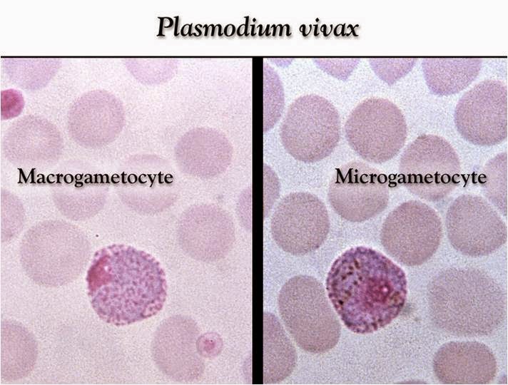
Male gametocyte(Microgametocyte)
Oval in shape
Diffused nucleus in centre
Diffused Pigments
Female gametocyte(Macrogametocyte)
Oval in shape
Compact nucleus in
corner
Diagnostic points
ØRed cells containing
parasites are usually enlarged.
ØSchuffner's dots are frequently
present in the red cells as shown above.
ØThe mature ring forms
tend to be large and coarse.
ØDeveloping forms are
frequently present.

Plasmodium falciparum
Early trophozoite
(ring form)

Ø1or
2 red nuclei on the ring-like light blue cytoplasm ; multiple infection in a
cell.
ØInfected RBCs normal in size
Male gametocyte (Microgametocyte)

ØBanana
shaped
ØDiffused
nucleus in the centre
ØMalarial
pigment diffused
Female gametocyte (Macrogametocyte)

ØCrescent
shaped
ØCompact
nucleus in centre
Diagnostic points
ØRed
Cells
are not enlarged.
ØRings
appear fine and delicate and there may be several in one cell.
ØSome
rings
may have two chromatin dots.
ØPresence
of marginal or applique forms.
ØIt
is unusual to see developing forms in peripheral blood films.
ØGametocytes
have a characteristic crescent shape appearance.
However, they do not usually appear in the blood for the first four weeks of infection
However, they do not usually appear in the blood for the first four weeks of infection
ØMaurer's
dots may be present.
 |
| Differentiating characteristics of Plasmodium sp. |
BY:
ESWARI VASANTH
SENIOR TECHNICIAN
ROHFW, CHENNAI-90
No comments:
Post a Comment